Prostate Biopsy Pathology
Prostate core biopsy read: cancer per core, Gleason and ISUP grade, % pattern 4, tumour length.
Pelvis4 sections
WSI· wsi
Thyroid FNA read: adequacy, Bethesda category I–VI, risk of malignancy and usual management.
This model is not available to run yet. Vote to show interest and get an email when it opens.
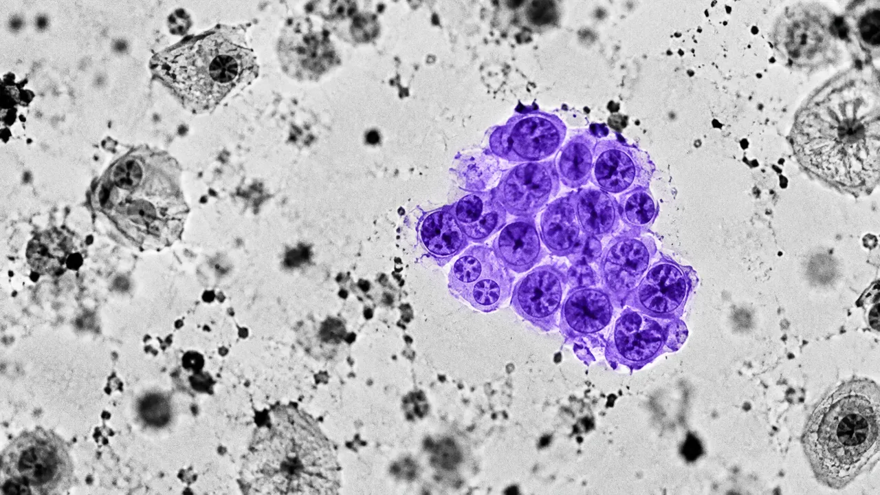
Thyroid FNA Cytology reads a scanned thyroid fine-needle aspiration, either a direct smear or a liquid-based preparation. Fine-needle aspiration is the step after a suspicious nodule on ultrasound, and its category decides between follow-up, repeat aspiration, molecular testing and surgery.
The run assesses adequacy and assigns a category of The Bethesda System for Reporting Thyroid Cytopathology, 3rd edition (TBSRTC 2023), from I to VI. With the category it returns the risk of malignancy that TBSRTC 2023 associates with it and the usual management, so the result reads the way a cytology report is used at the endocrine clinic.
Decision support for the cytopathologist, with most value in the indeterminate categories III (atypia of undetermined significance) and IV (follicular neoplasm), where reader agreement is lowest and the next step is least obvious. For these categories the guidance lists the options TBSRTC 2023 gives, such as repeat aspiration, molecular testing or diagnostic lobectomy, without choosing between them.
Cytopathologists, general pathologists who report thyroid FNA without subspecialty training, and endocrinologists or surgeons reviewing outside cytology. Developers use the run to close the loop between a thyroid ultrasound report and its cytology result in one data model.
| Requirement | Why |
|---|---|
| Stain recorded with the slide | Pap and Romanowsky stains show nuclear and colloid features differently |
| Pixel spacing in µm/px | Nuclear size and follicular-group size are compared in micrometres |
| Focus through thick smear areas | Follicular groups and papillary fragments are three-dimensional |
The ultrasound finding (TI-RADS category, nodule size and location) and prior cytology can be passed in
clinical_context, where they inform the management guidance.
| Section | Content |
|---|---|
| Bethesda category | Adequacy, TBSRTC 2023 category I–VI, the associated risk of malignancy and usual management |
| Draft report | Opt-in (options.report): an English narrative that interprets the result for the reader (findings, impression, limitations); every number is checked against the findings |
DiagnosticReport with an Observation for the category.| Category | Name |
|---|---|
| I | Nondiagnostic |
| II | Benign |
| III | Atypia of undetermined significance (AUS) |
| IV | Follicular neoplasm |
| V | Suspicious for malignancy |
| VI | Malignant |
Thyroid Ultrasound (us/thyroid) grades the nodule with ACR TI-RADS and recommends aspiration; this model reads the
resulting cytology, linking imaging risk to the cytology category for the same nodule.
One run returns every section its input supports.
Prostate Biopsy Pathology
Prostate core biopsy read: cancer per core, Gleason and ISUP grade, % pattern 4, tumour length.
Any-Organ H&E Slide Analysis
Organ-agnostic H&E read: slide QC, tumour map, nuclei and mitoses, primary-site ranking, embeddings.
Breast Pathology
Breast case read from H&E, IHC and node slides: type, grade, biomarker scores, TILs and pN.
Colorectal Pathology
Colorectal polyp and cancer read: adenoma or serrated histology, dysplasia, MSI, tissue map.
Lung Cancer Pathology
Lung biopsy and resection read: NSCLC subtype, IASLC grade, PD-L1 TPS and EGFR mutation probability.
Peripheral Blood Smear
Peripheral blood smear read: classified white cells, differential percentages and a blast alert.