Coronary Angiography
Coronary angiography read: vessels, SYNTAX segments and stenosis with QCA evidence.
Cardiac13 sections
XA· cinefrom $0.12 / request
Live models first, then the coming-soon models with the most votes.
110 models · 1 live
Coronary Angiography
Coronary angiography read: vessels, SYNTAX segments and stenosis with QCA evidence.
Chest CT
Chest CT read: nodules with Fleischner or Lung-RADS, PE with RV strain, lungs, pleura and ribs.
Chest X-ray
Chest radiograph read: urgent flags, findings with boxes, device tips, CTR and interval change.
Colour Fundus Photography
Fundus photo read: DR grade, glaucoma suspicion, AMD stage, vessels and referral urgency per eye.
DICOM Tools
Inspect a DICOM study, convert it to NIfTI, PNG or MP4, and validate DICOM objects.
Echocardiography (TTE)
Full TTE read: views, LVEF, chambers, diastology, valves and effusion as ASE measurements.
Head CT
Head CT read: bleed volumes, ASPECTS, vessel occlusion, perfusion core and skull fractures.
Musculoskeletal X-ray
Bone and joint radiograph read: fractures, dislocations, bone age, osteoarthritis and alignment.
Obstetric Ultrasound
Trimester-aware pregnancy scan read: planes, biometry, GA, EFW, centiles, fluid and Doppler.
Radiology Report Tools
Report text tools: narrative from findings, structuring, error check, coding, simplify, translate.
Resting 12-Lead ECG
12-lead ECG read from signal, PDF or photo: rhythm, QTc, STEMI-equivalents and AI-ECG screens.
Thyroid and Neck Ultrasound
Thyroid nodule list with ACR TI-RADS level and FNA advice, gland volume and cervical lymph nodes.
3D Export & Quantification
Turn segmentations into STL and OBJ meshes, 3D renders and rotating videos, and volume measurements.
Abdomen & Pelvis CT
Abdomen-pelvis CT read: acute abdomen flags, organ lesions with management, stones and trauma.
Abdominal and Renal Ultrasound
Organ-by-organ abdominal read: liver, gallbladder, kidneys, spleen, aorta and paediatric bowel.
Brain MRI
Brain MRI read: stroke, tumours, MS lesions, aneurysms, atrophy and ARIA in one result.
Brain PET and DaT SPECT
Brain PET and DaT SPECT read: Centiloid, CenTauRz, FDG dementia patterns and striatal binding.
Breast Ultrasound
Breast lesion list with BI-RADS v2025 descriptors and final assessment, axillary nodes and ABUS.
Cardiac CT
Cardiac CT read: Agatston calcium, CAD-RADS 2.0 stenosis, plaque, chambers and TAVI measurements.
Clinical Skin Photo (rash or lesion)
Rash and lesion photo read: top-5 differential, urgency, photo quality and severity scores.
Colonoscopy
Recorded colonoscopy read: polyps with optical diagnosis, BBPS, caecal intubation, withdrawal time.
Dental CBCT
CBCT read: segmented teeth, jaws and nerve canals, third-molar canal relations and airway size.
Dermoscopy · Skin Lesion
Dermoscopic lesion read: ranked differential, malignancy risk, structures and 7-point score.
eFAST Trauma Ultrasound
eFAST read per window: free fluid, pericardial effusion, pneumothorax and an overall result.
FDG PET/CT (Oncology)
Oncologic FDG PET/CT read: lesions with SUV, Lugano stage, Deauville score and metabolic response.
Gynaecological Pelvic Ultrasound
Gynaecological pelvic read: O-RADS adnexal masses, endometrium, uterus and fibroids, follicles.
Holter & Ambulatory ECG
Ambulatory ECG read: arrhythmia episodes, ectopy and AF burden, pauses, HRV and evidence strips.
Image Registration
Align two series or studies: rigid, affine or deformable transforms with resampled images and QA.
Lung Ultrasound
Per-zone lung ultrasound read: B-lines, sliding, consolidation, effusion, BLUE profile, LUS score.
Mammogram (2D)
Mammogram read: suspicion score, recall, BI-RADS, density, located lesions and risk.
Panoramic Radiograph (OPG)
OPG read: FDI-numbered odontogram, caries, periapical lesions, third molars and bone level.
Promptable Segmentation
Outline any structure in a medical image: click, box or scribble, name it, or label every organ.
Prostate Biopsy Pathology
Prostate core biopsy read: cancer per core, Gleason and ISUP grade, % pattern 4, tumour length.
Prostate MRI
Prostate MRI read: PI-RADS v2.1 lesions, sector map, PSA density and fusion-biopsy targets.
Retinal OCT
Retinal OCT read: layers, fluid volumes, AMD, DME, RNFL and change since the last visit.
Spine CT
Spine CT read: every vertebra labelled, fractures with AO Spine type, alignment and canal size.
Spine MRI
Spine MRI read: a graded level table, cord measurements and red flags in Fardon 2014 terms.
Vascular Duplex Ultrasound
Duplex read: carotid stenosis and IMT, DVT compression and peripheral arterial waveforms.
Abdominal MRI
Abdominal MRI read: liver fat and iron, LI-RADS, MRCP, pancreatic cysts, Bosniak and kidney volume.
Abdominal X-ray
Abdominal film read: bowel gas pattern, free air, radio-opaque stones and swallowed objects.
Any-Organ H&E Slide Analysis
Organ-agnostic H&E read: slide QC, tumour map, nuclei and mitoses, primary-site ranking, embeddings.
Bone Scintigraphy
Whole-body bone scan read: metastatic hotspots, Bone Scan Index, PCWG3 progression and ATTR-CM flag.
Breast MRI
Breast MRI read: enhancing lesions, kinetics, BI-RADS assessment, BPE, FGT and implant integrity.
Breast Pathology
Breast case read from H&E, IHC and node slides: type, grade, biomarker scores, TILs and pN.
Breast Tomosynthesis
DBT read: lesions located in 3D with their best slice, exam score, recall and BI-RADS.
Capsule Endoscopy
Capsule study read: bleeding sources, lesions with Saurin relevance, transit times and key frames.
Cardiac MRI
Cardiac MRI read: ventricular function, scar, T1/T2/ECV mapping, flow and strain per AHA segment.
Cerebral DSA
Cerebral angiogram read: vessels, aneurysms, AV shunts, occlusions, stenosis and eTICI grading.
Colorectal Pathology
Colorectal polyp and cancer read: adenoma or serrated histology, dysplasia, MSI, tissue map.
CT Angiography (Aorta, Carotid, Run-off)
Arterial CTA read: aortic diameters, dissection and aneurysm, endoleak, carotid and leg stenoses.
Head & Neck CT
Neck, sinus and temporal-bone CT read: nodes by level, Lund-Mackay, airway, abscess and tumour.
Image Embeddings and Similar Cases
Medical image embeddings for search, clustering and zero-shot labels, plus similar-case retrieval.
Intra-oral Radiographs (Bitewing and Periapical)
Bitewing and periapical read: surface caries depth, bone levels, PAI and restorations on one chart.
Intravascular OCT
IVOCT pullback read: lumen, plaque phenotype, calcium, stent sizing and strut-level stent analysis.
Intravascular Ultrasound (IVUS)
Vendor-neutral IVUS pullback read: lumen and EEM, plaque burden, calcium, sizing and stent result.
Knee MRI
Knee MRI read: ACL status, meniscal tears, cartilage defects and bone marrow oedema.
Lateral Cephalogram
Ceph tracing: landmarks, cephalometric analyses, skeletal class and CVM growth stage.
Lung Cancer Pathology
Lung biopsy and resection read: NSCLC subtype, IASLC grade, PD-L1 TPS and EGFR mutation probability.
Medical Image Q&A
Ask any question about a medical image: draft answers with grounding boxes and prior comparison.
Musculoskeletal CT
Pelvis and extremity CT read: fractures with fragments, 3D bone models and Bone-RADS lesions.
Musculoskeletal Ultrasound
Region-aware MSK read: rotator cuff, carpal tunnel, synovitis, infant hip (Graf) and abscess.
NICU Radiograph
Neonatal babygram read: umbilical line and tube tips by vertebral level, plus NEC signs.
Nuclear Cardiology (MPI and Cardiac Amyloid)
Nuclear cardiology read: perfusion scores, gated function, myocardial blood flow and ATTR-CM grade.
Oncology Staging & Response CT
Staging and response CT: lesion inventory, RECIST 1.1 table, tracked lesions and Node-RADS nodes.
Opportunistic CT Biomarkers
Biomarker panel from routine CT: sarcopenia, bone density, liver fat, aortic calcium, fractures.
Peripheral Blood Smear
Peripheral blood smear read: classified white cells, differential percentages and a blast alert.
PSMA PET/CT
PSMA PET/CT read: PROMISE V2 miTNM staging, PSMA tumour volume and Lu-PSMA eligibility signals.
Radiotherapy Planning CT
Planning CT contouring: site-specific organs at risk and a GTV proposal as a DICOM RTSTRUCT.
Spine X-ray
Spine radiograph read: scoliosis curves, vertebral fractures, sagittal balance and slip grades.
Upper GI Endoscopy (EGD)
Gastroscopy read: station completeness, LA grade, Barrett's extent and gastric neoplasia flags.
Wearable & Single-Lead ECG
Smartwatch and handheld ECG strips: rhythm, AF, heart rate, QRS and QTc, and strip quality.
Wound & Burn Photo
Wound and burn photo read: area in cm², tissue types, staging, foot flags and healing trend.
Anterior Segment Imaging
Anterior segment read: keratitis triage, cataract grade, keratoconus stage and angle closure.
Bone Marrow Aspirate
Marrow aspirate read: cell-by-cell myelogram, blast percentage, M:E ratio and dysplasia flags.
Bronchoscopy
Recorded bronchoscopy read: endobronchial tumours and mucosal abnormalities, located by airway.
Cath-Lab Hemodynamics
Cath pressure tracings read: FFR and pullback pattern, valve gradients, right-heart values, QA.
Cervical Cytology (Pap Test)
Pap test read: adequacy, Bethesda 2014 category and a ranked gallery of abnormal cells.
Contrast-Enhanced Mammography
CEM read: enhancing masses and non-mass enhancement, BPE level and BI-RADS by the CEM lexicon.
Cystoscopy
Recorded cystoscopy read: papillary tumours and suspicious flat lesions, mapped with key frames.
DXA (Bone Densitometry)
DXA read: T-scores by ISCD rules, VFA fractures, aortic calcification and body composition.
Exercise Stress ECG
Treadmill and bicycle stress ECG read: ST response by stage, arrhythmias and Duke Treadmill Score.
Fetal MRI
Fetal brain MRI read: 3D reconstruction, tissue volumes and biometry against gestational age.
Heart Sounds (Digital Stethoscope)
Digital stethoscope recordings read: S1/S2, murmur presence, timing and grade, echo referral risk.
Hysterosalpingogram
HSG read: fill and spill for each fallopian tube, with occlusion level and hydrosalpinx.
Intra-oral 3D Scan
Intra-oral scan processing: FDI-labelled tooth segmentation, missing teeth and dental landmarks.
Joint MRI
Joint MRI read: rotator cuff tears, retraction and fatty infiltration, bone and muscle masks.
Laparoscopic Surgery Video
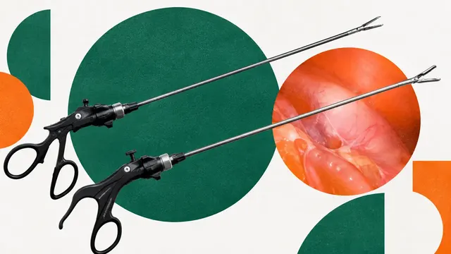
Recorded laparoscopy review: phase timeline, critical view of safety, instruments and anatomy masks.
Laryngoscopy and Stroboscopy
Laryngoscopy and stroboscopy read: vocal-fold lesions and glottal-area segmentation with waveform.
Lung V/Q Scintigraphy
Lung V/Q read: segmental mismatched perfusion defects and a pulmonary embolism call per EANM 2019.
LV Ventriculography & Aortography
LV gram and aortogram read: volumes, LVEF, regional wall motion and Sellers regurgitation grades.
Neonatal Cranial Ultrasound
Preterm cranial ultrasound read: Papile IVH grade, ventricular index and dilatation flag.
Nerve Block Ultrasound
Recorded nerve-block clip read: nerves, vessels, fascia, pleura and needle tip for QA and teaching.
OCT Angiography
OCTA read: foveal avascular zone, vessel and perfusion density, non-perfusion and new vessels.
Ocular Ultrasound (POCUS)
Ocular POCUS read: optic nerve sheath diameter and retinal detachment versus vitreous disease.
Pelvic MRI
Pelvic MRI read: rectal cancer staging, gynaecological staging, O-RADS MRI, fibroids and fistulas.
Peripheral & Visceral Angiography
Peripheral and visceral DSA read: patency, stenoses, runoff, TASC II and GLASS stage, extravasation.
Prostate Ultrasound (TRUS and Micro-US)
Prostate read: gland outline and volume, PSA density and suspicious lesions on micro-ultrasound.
Renal Scintigraphy
Renal scintigraphy read: diuretic renogram curves, split function, drainage and DMSA cortical scars.
Retinopathy of Prematurity Screening
ROP read: plus disease, ICROP3 zone and stage, A-ROP and treatment-warranted ROP per eye.
Scrotal Ultrasound
Scrotal read: torsion signs on Doppler, intratesticular masses and microlithiasis.
Somatostatin Receptor PET/CT
SSTR PET/CT read for neuroendocrine tumours: lesions, SSTR-RADS 1.0 and Krenning score.
Swallow Study (VFSS)
Videofluoroscopic swallow read: PAS per bolus, aspiration frames, hyoid and UES motion, residue.
Theranostics SPECT/CT Dosimetry
Post-therapy Lu-177 SPECT/CT: organs at risk, time-activity curves and absorbed dose per cycle.
Thyroid FNA Cytology
Thyroid FNA read: adequacy, Bethesda category I–VI, risk of malignancy and usual management.
Thyroid Scintigraphy
Thyroid scintigraphy read: uptake pattern, thyroid uptake percentage and hot or cold nodules.
Total-Body Photography (mole mapping)
Total-body photo read: every lesion mapped, the most suspicious ranked, new and changed flagged.
Transoesophageal Echocardiography (TEE)
TEE read: 28 standard views, LAA thrombus, mitral valve anatomy and endocarditis signs.
Urine Cytology
Urine cytology read: Paris System 2.0 category, N/C ratio per cell and a gallery of atypical cells.
Voiding Cystourethrogram
Voiding cystourethrogram read: reflux side and International Reflux Study grade I–V.
Whole-body MRI
Whole-body MRI read: myeloma marrow infiltration and bone metastasis burden, with response.